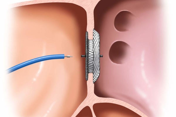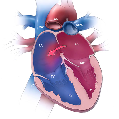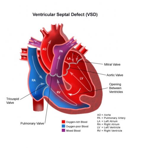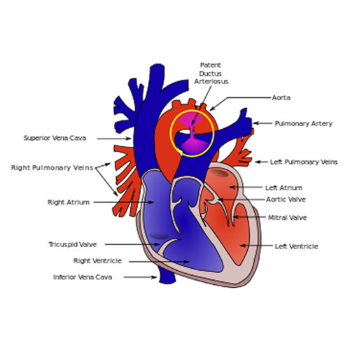
Device Closure is used to close a defect or an opening between the right and left sides of the heart. A heart has four chambers. The upper two chambers are called at Atria. Atria are separated by a wall which is called Atrial Septum. The lower chambers are known as Ventricles- Right and left. Pulmonary Veins supply Oxygenated blood to the Left Atrium. This left atrium, in turn, sends this oxygenated blood to the Left Ventricle through Mitral Valve, for circulation of oxygenated blood in the body.
The walls of the heart are well separated maintaining a good circulation system. But in some congenital heart defect situations, if there are any holes on the heart, either in the atrial wall or the ventricle wall, this can result in disturbed blood circulation resulting in performance failure of the heart. If the holes are particularly small, they can be filled up surgically, with some metal parts. This concept is known as DEVICE CLOSURE. So, in short, in Device Closure, there is a metal device that is surgically placed in the place where there is a whole in a heart, in order to maintain the heart’s performance.
What is Device Closure Procedure?
PROCEDURE OVERVIEW
The device closure procedure used as a reparative surgery is known as transcatheter closure, although it is done for a reparable hole and this is not the only treatment for hole closures. When compared to other surgeries, it is a less invasive surgery.
DURING THE SURGERY:
Before the surgery starts, a patient is administered with IV and intravenous medicines are started. The person has leads attached to his chest for ECG. The person’s groin/arm area is cleaned and a plastic thin tube called a catheter will be inserted through that area. Meanwhile, the patient is given sedatives for relaxation purposes and local anesthesia, but they will be awake and conscious throughout the procedure.
A contrast dye would be injected in the arteries so as to get an idea of the heart chambers and the size of the hole. If required, an echocardiogram or transoesophageal echocardiogram is also taken.
After the size of the hole is determined, a special catheter is administered in the arteries which carry a small metallic mesh-like closing device. This device is then fitted in the hole and then released by the catheter. This catheter is then taken out, eventually, heart tissue develops on the closure device, thus making it a part of the heart itself.
POST SURGERY:
When the catheter is taken out, suturing of the area is done or heavy bandaging, as required. The patient is kept in the hospital for a day or two. The patient can resume their routine a week after the doctor’s advice. They are advised to maintain a healthy lifestyle which includes exercising, maintaining a good diet, and regular cardiac checkups.


